Showing posts with label cancer. Show all posts

Wednesday, 4 July 2012

Under your skin: taking skin cancer out by its roots

the sun can cause field cancerization
Tumours can spring up amongst
'fields' of healthy skin cells.
The summer sun may finally be on its way. This is great news for barbecue kings and beach bums but also for the weeds lurking below the surface of the soil popping up intermittently to strangle my carrots.

New research published in Cell describes another reason to cake ourselves in sun cream, cover up bare flesh and wear ridiculously wide-brimmed hats in the coming months: weed-like skin cancers which start below the surface of the skin and grow upwards.

"Too much sun" is well known to carry a risk of skin cancer. Prolonged exposure to the sun's Ultraviolet (UV) rays can wither the DNA in cells on the skin's surface sometimes causing multiple tumours to spring up at once.

Dr Bing Hu and colleagues found alarming evidence that some cancers may start much deeper in the tissue. This may explain why skin cancers frequently reappear: surgery may remove a tumour, but its roots may remain.

The new research suggests that skin cancer can be kick-started by changes in the dermis – deeper skin tissue where healthy cells reproduce to replenish the cells on the surface.

the roots of skin cancers can begin deep in the tissue
The root cause of certain skin
cancers may be much deeper in the
tissue than previously thought.
The team, from the University of Lausanne in Switzerland, discovered that UV light can cause mutations in the wiring of dermal cells, specifically to a protein called 'Notch' which is necessary for individual skin cells to communicate.

They found that mice born with malfunctioning Notch develop severely distorted skin full of lesions and tears. The dermal skin cells of these mice displayed accelerated cell division, a common prelude to tumour formation.

The mouse studies gave the team a vital clue of what to look for in human cells. They have since discovered Notch is disrupted in some human skin cancers too.

Dr Hu says that designing drugs to protect or repair Notch in human cells might be used in “preventing or reversing” the unseen effects of the sun on cells under our skin.

Until then, whether you're weeding, barbe-ing or bathing this summer the advice remains the same: enjoy responsibly. 

And remember to buy ice. And fire-lighters.

Reference:

Hu B, Castillo E, Harewood L, Ostano P, Reymond A, Dummer R, Raffoul W, Hoetzenecker W, Hofbauer GF, & Dotto GP (2012). Multifocal Epithelial Tumors and Field Cancerization from Loss of Mesenchymal CSL Signaling. Cell, 149 (6), 1207-20 PMID: 22682244
ResearchBlogging.org

Tuesday, 19 June 2012

Who needs NASA? Launching genes with lasers in space-travelled fish

promoters launch genes on DNA
Inside the cell genes are launched from promoters on our DNA.
(photo of space shuttle Atlantis)
NASA has its sights on launching rockets into space using lasers. "What if..." they're wondering, "shuttles could be sent up using laser beams to heat their fuel from the ground?"

Biophysicists in Japan have had a similar idea. They've successfully used lasers to 'launch' genes inside living creatures, with a little help from nanotechnology. If this process works in humans, future battles with cancer may be fought by remote control.

Deep within our cells, genes are launched into action from promoters, sequences of DNA where movable machinery assembles to fire copies of a gene, called messenger RNAs (mRNAs), from the nucleus to the cytoplasm.

Promoter ’launch pads’ are triggered by different things – stresses or chemicals or signals from outside the cell. Some promoters are heat sensitive, firing off mRNAs in response to fever or infection. Arriving in the cytoplasm, their mission is to build proteins to defend the cell from invaders such as viruses.

New research published recently in PNAS, describes a way of using laser light to trigger these 'heat shock' promoters from above the skin of living organisms. It's a first step towards launching our own genetic defences to disease from outside the human body.
laser-fired genes may fight cancer
Could lasers be used to fire mRNAs out from
the nucleus to fight diseases?
(photo: Space shuttle Atlantis from plane, Ryan Graff)

To develop these new pyrotechnics, Eiliro Miyako and colleagues injected carbon nanoparticles called nanohorns into medaka fish, Oryzias latipes. These fish are no strangers to laboratories. They've even been to space (and were the first Earthly vertebrates to reproduce in orbit).

Nanohorns, molecule-thin sheets of carbon folded into cone shapes, have huge potential for scooping up and delivering drugs inside cells and tissues. But it was something else about these tiny metal structures (which measure around 1/100000 cm across) that excited Dr Miyako and his team: nanohorns convert laser light energy into heat.

With a microscopic fuel source in hand the team from collaborating research institutes in Japan, set about building a DNA launch pad.

They pieced together DNA in the lab, placing the gene for a green fluorescent protein (GFP) next to a man-made heat shock promoter. After transferring the whole thing into the cells of the medaka fish, it was time for launch!

A low-powered laser beam was focused beneath the fish's skin. The carbon nanohorns absorbed the laser's energy, emitting it as heat. The surrounding tissue began to warm up. At a temperature of 42°C the heat shock promoters fired into life, launching the GFP gene. Minutes later the cells in the fish were glowing green. Genes had been successfully launched from outside a living body.

remote control gene expression
Medaka, the first vertebrate to reproduce in space.
In this study its genes were launched by remote control.
Dr Miyako writes "This work is a proof-of-principle study demonstrating that... gene expression can be mediated by the photothermal properties of nanocarbons."

 He believes that they could be used "in various biological fields, including analysis of cell signaling within organisms, investigation of genetic mechanisms, and development of unique cell therapies and tissue engineering techniques."


Makes you wonder which will come first - the fire laser-propelled rocket to the moon, or the first cancerous cell killed by remote control?

What does this mean for me?
This study was not simply about making glowing fish. As Dr Miyato says, this is a proof of principle. In the future, lasers might be used to trigger specific genes inside the human body, boosting the body's response to infection, or triggering cell death in cancer cells. This could compliment drug-based approaches aiming to manipulate genes and proteins in a similar way. The team have also used nanohorns to trigger genes inside living mice and found no signs of toxicity or adverse reaction to the particles, which is encouraging for future trials.

What does this mean for science?
Remote control of gene expression has been achieved before, but this study is the first to use near infrared light (NIR, with wavelengths between 0.7- 2.5um). NIR light lies inside the "optical window" of biological tissue (0.6- 1.1um) and is able to penetrate over 10cm deep. This study adds to the - already impressive - list of potential uses for metal nanoparticles in biology including drug delivery, tissue scaffolding,  the detection of harmful pathogens and improved MRI images.


Reference (free to download via Open Access!):

ResearchBlogging.org Miyako, E., Deguchi, T., Nakajima, Y., Yudasaka, M., Hagihara, Y., Horie, M., Shichiri, M., Higuchi, Y., Yamashita, F., Hashida, M., Shigeri, Y., Yoshida, Y., & Iijima, S. (2012). Photothermic regulation of gene expression triggered by laser-induced carbon nanohorns Proceedings of the National Academy of Sciences, 109 (19), 7523-7528 DOI: 10.1073/pnas.1204391109

Thursday, 7 June 2012

Death by metal: a hidden detonator inside cancer cells?

Our cells are wired to explode. Given the right signals they can burst open, scattering bits of crunched up DNA, shrivelled membrane and chemicals in all directions. Sometimes this is all part of the plan: controlled cell death it vital to defining the outline of our toes and fingers in the womb, and to the daily act of replacing old cells with new ones. Cell death is a part of life.

pools of iron are found in some cancer cells
Could pools of iron inside cancer cells be exploited
to trigger their demise?
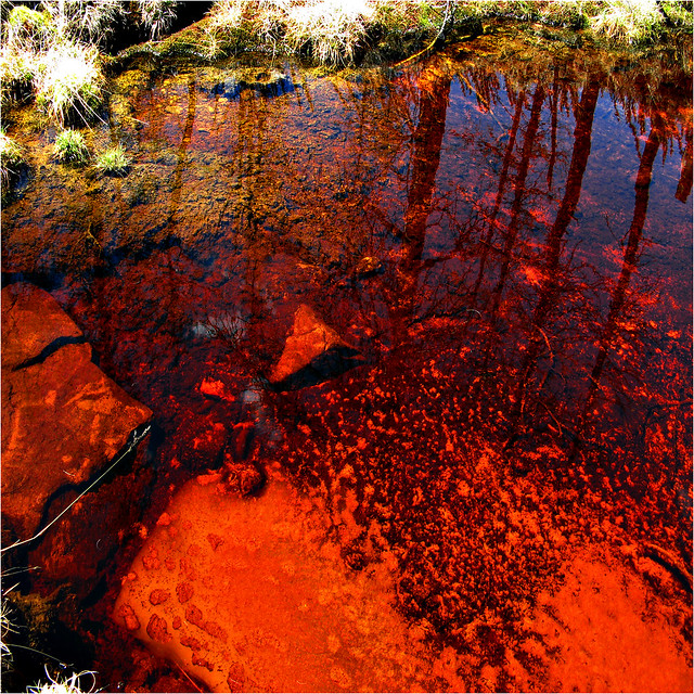
Iron in Lake Khövsgöl, Mongolia
(picture credit Josefontheroad
New research has uncovered a hidden route to cell death. Death by iron, or 'ferroptosis' may be a secret weapon against some forms of cancer.

In work published recently in Cell, Scott Dixon and colleagues triggered the death  of cells in a dish using chemicals which causes a build-up of Reactive Oxygen Species (ROS). ROS are volatile and highly damaging to cells, so death within a few hours came as no surprise. What did was another observation: erastin was only effective in cells with a healthy supply of iron.

Iron absorbed from the blood stream (but not other heavy metals such as copper, nickel or cobalt) appeared to sensitise certain cells to erastin and a quick death. 

Exactly what the link is between ROS-inducing chemicals such as erastin and iron has yet to be uncovered. But the team from Columbia University, New York, found evidence that ferroptosis has a "unique genetic network" that is entirely separate from other forms of cell death such as apoptosis (the 'culling' of cells, apoptosis helped to create the gaps between our toes) and necrosis (triggered when a cell is too injured to repair).


This distinct wiring presents an intriguing opportunity: to selectively activate ferroptosis to kill certain cancer cells.

Death by Iron, targeting Ferroptosis
Can death by iron, or 'ferroptosis' be aimed at cancer?
Or blocked in nerve cells to protect the nervous system?
(Iron Maiden. ' The Trooper' (1983))
"The RAS family [of genes] is mutated in 30% of cancers," Dr Dixon writes. These mutations lead to uncontrolled cell division, but also "for better or worse... elevated levels of iron... are observed in some cancer cells".

His team believes it is possible to activate ferroptosis in RAS-mutated cancers inside the human body, using the abnormal iron levels to sensitise the cells to chemicals like erastin.

But activating ferroptosis in cancer may not be its only health benefit. There may be a use for blocking the process too.

The team successfully rescued neurons in rodent brains from cell death by blocking ferroptosis with a chemical inhibitor called ferrostatin-1. They propose that blocking ferroptosis in human brain cells following a stroke or epileptic fit (when ROS and iron levels are high) might protect the central nervous system from long-term damage.

Although the wiring inside our cells is complex (and multitasking is common), ferroptosis is a rare example of independence. Its distinct wiring may allow selective activation or inhibition of cell death, and maybe even the treatment of cancer with fewer side effects. That this metal-based killer might be used to protect life is, in more ways than one, quite ironic.

What does this mean for me?
This study might lead to a whole new line of approach for the treatment of some cancers and diseases which damage the central nervous system. High levels of iron have been reported in cases of Alzheimer's and Parkinson's disease. Understanding exactly how our cells are wired to use ferroptosis will make it easier for scientists to manipulate its effects with drugs similar to erastin or ferrostatin-1.

What does this mean for science?
The discovery of a previously unknown route to cell death shows just how much about our cells we have yet to understand. Indeed, the authors of this work suggest there may be much more "hidden" wiring  used by the cell, waiting to be discovered.

Reference:

ResearchBlogging.orgDixon, S., Lemberg, K., Lamprecht, M., Skouta, R., Zaitsev, E., Gleason, C., Patel, D., Bauer, A., Cantley, A., Yang, W., Morrison, B., & Stockwell, B. (2012). Ferroptosis: An Iron-Dependent Form of Nonapoptotic Cell Death Cell, 149 (5), 1060-1072 DOI: 10.1016/j.cell.2012.03.042

Thursday, 17 May 2012

Anchors away! When neural stem cells decide a change is as good as a rest

neural stem cells in a niche
Neural stem cells are anchored to their niche until they
decide to migrate.
Painting: 'Canada Timber Docks, Liverpool.
Towards close of day' by Robert Dudley (active 1865-1891)
Between 1830 and 1930, over nine million people left England from Liverpool  on ships bound for Australia, Canada and America. The Merseyside port swelled with would-be emigrants, all holding tightly to the decision to leave their homes for the promise of a new life.

Stem cells in the brain are similarly destined for change. A recent study suggests their transformation into specialised cells, a process known as differentiation, is combined with the decision to migrate to where they are needed, bringing new understanding of the development and repair of brain tissue.

In the brain, neural stem cells (NSCs) can be found anchored in  'niches': port-like microenvironments which shelter the cells in a dormant, undifferentiated state. NSCs might eventually migrate all over the brain, some becoming neurons along the way, but this can only happen correctly if differentiation is timed precisely with release from the niche.

In a paper published recently in Nature Cell BIology, Francesco Niola and colleagues found the same set of proteins inside a neural stem controls both anchorage to the niche and the onset of differentiation, synchronising the two processes. This control may prevent differentiation from misfiring, leading to problems in development or even cancer.
stem cell migration
Neural stem cells migrate to different parts of the brain, becoming different
types of brain cell such as neurons.
Painting: 'Ship off Liverpool', Robert Salmon (1811)

The team from Colombia University, New York looked inside mouse NSCs. They found that Inhibitor of DNA-binding (Id) proteins prevent an NSC from differentiating too soon by repressing the transcription of certain genes. Id proteins were also found to control RAP1, a protein involved in adhesion between the stem cell and its niche.

Dr Anna Lasorella, a senior author of this paper said a key question for the future was to, "determine whether Id proteins also maintain stem cell properties in cancer stem cells in the brain." "In fact," she said, "normal stem cells and cancer stem cells share properties and functions." She added that targeting Id proteins in cancer stem cells might, "lead to more effective therapies for malignant brain tumours".


What does this mean for me?
Better understanding of how neural (and other) stem cells differentiate may influence when and where injected stem cell therapies are used. Also, as Dr Lasorella said (above), studying processes involved in stem cell regulation may give insight into similar processes in cancerous stem cells leading to malignant brain tumours.


What does this mean for science?
This study presents a new idea - it was previously thought that the niche itself controls the release of NSCs with chemical signals. Here we see the decision is influenced, at least in part, by the NSC's internal wiring. More generally, the central role of Id proteins is another good example of multi-tasking, involving co-ordination between internal wiring of differentiation and the cell's external environment.


nature cell biologyReference:

ResearchBlogging.orgNiola, F., Zhao, X., Singh, D., Castano, A., Sullivan, R., Lauria, M., Nam, H., Zhuang, Y., Benezra, R., Di Bernardo, D., Iavarone, A., & Lasorella, A. (2012). Id proteins synchronize stemness and anchorage to the niche of neural stem cells Nature Cell Biology, 14 (5), 477-487 DOI: 10.1038/ncb2490

Friday, 11 May 2012

Faultless: Your skin's battle with open wounds and cancer

keratin repairs skin lesions
The San Andreas fault.
In our skin, fringes of cells meet to close a wound.
(picture credit: David Parker)
New research has revealed a connection between how our skin heals and the prevention of skin cancers.

In a paper published  two weeks ago in JCB, Jeremy Rotty and colleagues showed that keratin 6 (K6), a fibrous protein used to repair skin lesions, can also put the brakes on skin cells growing too quickly.

When the surface of your skin is scratched, the wound is quickly bridged by keratinocytes, cells full of K6 which migrate through the tissue and mesh together. Researchers found that K6, as well as providing scaffolding inside these cells, also attaches to and controls Src, a protein at the heart of the wiring for cell migration and growth.

It is here that a careful balance is struck. If there isn't enough Src activity inside a keratinocyte, it may not migrate at all, leaving wounds exposed. Too much Src, however, and the cells could migrate too far, growing into tumours. Skin cells lacking K6 to control Src activity have been shown to  produce aggressive cancers.
Keratinocytes migrate into a wound
Keratinocytes actually roll into a wound
in the skin, like stones into a valley.
(picture credit: moonjazz)

The researchers from John Hopkins University, Baltimore, USA suggested that inside skin cancer cells K6 might be a “protective mechanism that maintains epithelial (skin surface) tumours in a ... less aggressive state".


What does this mean for me?
Future treatments for skin cancer might target the relationship between K6 and Src. A lack of K6 might also be looked for as a marker for predisposition to certain types of skin cancer.


What does this mean for science?
This is a great example of multi-tasking inside our cells. Keratin K6 can regulate both the structure and movement of keratinocytes. The relationship between K6 and Src  shows how important the inner-wiring of each individual cell can be to the overall tissue, where lesions need to be repaired despite the risk of cancer.


JCB
Reference:
ResearchBlogging.org
Rotty, J., & Coulombe, P. (2012). A wound-induced keratin inhibits Src activity during keratinocyte migration and tissue repair The Journal of Cell Biology, 197 (3), 381-389 DOI: 10.1083/jcb.201107078


Tuesday, 27 March 2012

Too many live wires

Hello to anyone and everyone who has found themselves here! The idea behind this blog is to offer a fresh perspective on the complex life inside living cells. 
Tools of the trade: Pipette.
Used by biologists to mix precise
amounts of liquid such as DNA
in solution. (For hours on end.
Whilst tied to a lab bench.)

Why listen to me?

I am a systems biologist. I look inside cancer cells to examine the wiring between different genes and proteins which might be at fault. Then, because this wiring is often tangled, I get up from the microscope or lab bench and plonk myself behind a computer.

It is here that we use whatever we’ve been able to see to build a virtual model of parts of the cell. These models allow us to make sense of all of the information we see and, more importantly, to predict what might be happening to what we can’t see.

I’ll go into what Systems Biology actually is in a later post.


My posts will also try to answer questions like:

What does the latest “scientists find the gene for <insert something horrible here>” headline actually mean?

What’s in the research papers from around the world that most newspapers don’t report on?

and also…

How do I get into biology if I have a maths or computer science background?

and the tricky one... What are scientists actually like?

Wiring inside cancer cells: HeLa cervical cancer cells, enigneered in a dish to glow different colours
 as they prepare to go through cell division. We can learn a lot about the cell's inner wiring from
measuring how quickly these "traffic lights" change.
Watch the movie here!

I hope this blog will be unique, useful and <gulp> even entertaining! Any technical jargon will be explained, messages will hopefully be clear, and I won’t go on for pages and pages with some lofty opinion or other. I will hopefully be posting every two weeks (at least) and, unlike most blogs, there won’t be any recipes.

Apart from this one.

Tomato, caper and mint pasta sauce

(serves 4)
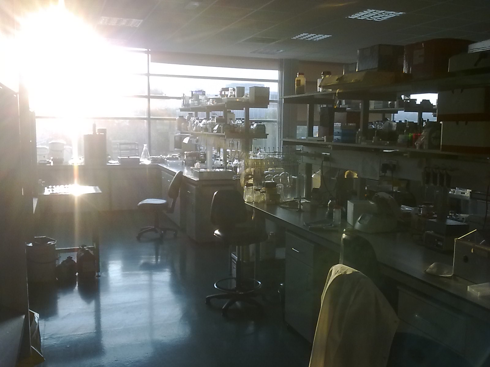
The lab. Science is a lot like cooking really.
Ingredients:

1 decent handful of spaghetti
2 tins of chopped tomatoes
1 large clove of garlic, crushed
2 tbsp olive oil
2 tbsp capers, drained
1/2 tbsp tomato puree
1 handful chopped mint
1 handful chopped basil
pinch of chili flakes
salt
pepper


Recipe:
Warm the olive oil in a pan over a medium heat, then add the garlic.
After 5 minutes or when garlic starts to brown, remove garlic and add tomatoes. Stir.
Simmer for 5 minutes then add puree and chili. Stir again.
Leave simmering for 10 minutes adding salt and pepper as you like.
Whilst this is bubbling away cook the pasta in salted water.
With one mintue to go add the capers, mint and basil. Stir well.
Drain pasta, serve and spoon over the sauce.

Enjoy!


Many thanks to Professor Mike White, Dr Dave Spiller and Rick Stein.